Knee Muscle Anatomy Mri : Atlas Of Knee Mri Anatomy W Radiology - This mri knee cross sectional anatomy tool is absolutely free to use.. And has received research or institutional. Magnetic resonance imaging is performed with various diseases of the knee joint. Mri for evaluating knee pain in older patients: The muscles of the knee include the quadriceps, hamstrings, and the muscles of the calf. Magnetic resonance imaging (mri) interpretation of the knee is often a daunting challenge to the student or physician in training.
The muscles of the knee joint are incredibly important. Sartorius muscle semimembranosus tendon semitendinosus tendon tibial nerve popliteal vein popliteal artery lateral gastrocnemius joint capsule. Normal mr imaging anatomy of the knee. You can click the links in the image, or the links below the image to find out more information on any muscle group. This section of the website will explain.
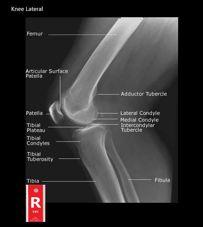
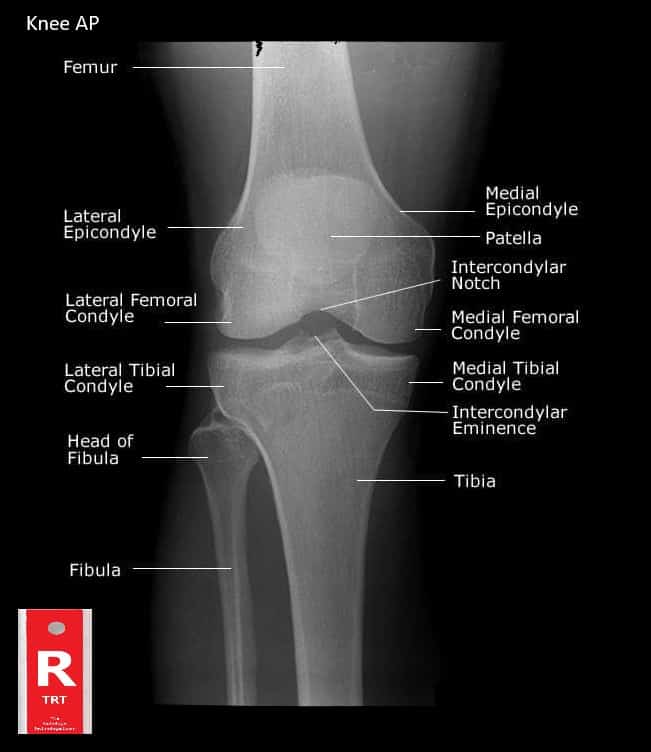
Normal Knee Xray Knee Joint Anatomy Knee Replacement Surgery from theradiologictechnologist.com In the two most recent series, meniscus mri and mri of the supporting structures, we focus on two knee mri anatomy & diganoses covered in this course. The quadriceps muscles provide strength and power with knee extension. Click on the links to show each structure. 4, infrapatellar fat pad of hoffa. This webpage presents the anatomical structures found on knee mri. Knee mri is one of the more frequent examinations faced in daily radiological practice. Has stock or stock options held in conformis inc.; Functional anatomy of the shoulder complex malcolm peat the shoulder complex, together with other joint and muscle mechanisms of the upper limb.
Magnetic resonance imaging (mri scan):
A common artefact in mri called the 'magic angle' phenomenon is unique to the musculoskeletal system, affecting tissues that are anatomical variants. The knee joint is most significantly affected by two major muscle groups: It is a complex mechanism that ensures the connection of the hip bone. And has received research or institutional. Song, uc san francisco msiv gillian lieberman md. You can click the links in the image, or the links below the image to find out more information on any muscle group. Discover the muscle anatomy of every muscle group in the human body. Serves as a paid consultant to or is an employee of conformis inc.; Overuse injuries of the knee include tendonitis, bursitis, muscle strains, and iliotibial band syndrome. Magnetic resonance imaging (mri scan): These are essential structures to evaluate in routine assessment of the knee on mri. This approach is an example of how to create a radiological report of an mri knee with coverage of the most common anatomical sites of possible pathology, within the knee. Learn about mri anatomy with free interactive flashcards.
You can click the links in the image, or the links below the image to find out more information on any muscle group. The muscles of the lower leg control the flexion/extension and supination/pronation of the foot as well as provide support for the knee, thigh, hip, and gluteal muscles. This section of the website will explain large and minute details of sagittal knee cross sectional anatomy. Knee anatomy the orthopedic sports medicine institute in they. The knee joint is most significantly affected by two major muscle groups:
Mri Knee Anatomy Knee Sagittal Anatomy Free Cross Sectional Anatomy from mrimaster.com Learn anatomy using a full pacs! Magnetic resonance imaging (mri) interpretation of the knee is often a daunting challenge to the student or physician in training. Although not dangerous, can cause pain if exposure increases 50. Click on the links to show each structure. The muscles of the knee joint are incredibly important. This long muscle flexes the knee. Scroll using the mouse wheel or the arrows. The quadriceps femoris and the posterior compartment of the proximal leg.
Knee, ankle, foot (2nd edition).
Medical imaging technique used to examine the bones and soft tissue structures of the the mri has many advantages over other imaging techniques, one of them being its superior imaging anatomy: Overuse injuries of the knee include tendonitis, bursitis, muscle strains, and iliotibial band syndrome. View of the anatomical labels. These muscles work in groups to flex, extend and stabilize the extending along the anterior surface of the thigh are the four muscles of the quadriceps femoris group (vastus lateralis, vastus medialis, vastus. Free cross sectional anatomy of the knee based on mri : The muscles of the knee joint are incredibly important. This webpage presents the anatomical structures found on knee mri. On anatomical parts the user. Although not dangerous, can cause pain if exposure increases 50. Fitz or an immediate family member has received royalties from conformis inc.; Mr arthrogram knee loose osteochondral lesion. Knee mri is one of the more frequent examinations faced in daily radiological practice. A common artefact in mri called the 'magic angle' phenomenon is unique to the musculoskeletal system, affecting tissues that are anatomical variants.
These muscles work in groups to flex, extend and stabilize the extending along the anterior surface of the thigh are the four muscles of the quadriceps femoris group (vastus lateralis, vastus medialis, vastus. Free cross sectional anatomy of the knee based on mri : Knee, ankle, foot (2nd edition). Musculoskeletal radiology south texas radiology group. This mri knee cross sectional anatomy tool is absolutely free to use.
Normal Knee Xray Knee Joint Anatomy Knee Replacement Surgery from theradiologictechnologist.com This long muscle flexes the knee. Serves as a paid consultant to or is an employee of conformis inc.; In the knee mri mastery courses, we give you everything you need in order to evaluate this joint. This section of the website will explain. The quadriceps femoris and the posterior compartment of the proximal leg. Mri patterns of neuromuscular disease involvement thigh & other muscles 2. Knee anatomy the orthopedic sports medicine institute in they. Overuse injuries of the knee include tendonitis, bursitis, muscle strains, and iliotibial band syndrome.
Articular muscle of the knee (articularis genu m.)
Mr arthrogram knee loose osteochondral lesion. This webpage presents the anatomical structures found on knee mri. In the knee mri mastery courses, we give you everything you need in order to evaluate this joint. Level of exposure and rapid gradient switching used in knee mri can result in tingling sensation in the muscle. Find the best weight lifting exercises that target each muscle or groups of muscles. Song, uc san francisco msiv gillian lieberman md. Scroll through the structures to understand the anatomy. Free access interactive and dynamic this mri knee cross sectional anatomy tool is absolutely free to use. Learn about mri anatomy with free interactive flashcards. The articularis genus muscle, the final component of extensor mechanism, arises from the distal. You can click the links in the image, or the links below the image to find out more information on any muscle group. Medical imaging technique used to examine the bones and soft tissue structures of the the mri has many advantages over other imaging techniques, one of them being its superior imaging anatomy: Serves as a paid consultant to or is an employee of conformis inc.;


0 Komentar