Kidney Blood Vessels Labeled / The renal artery is one of these two blood vessels.. But they are depicted in most pictures and labeled as segmental veins, or not labeled. The renal artery is one of these two blood vessels. These are the functional units. The renal artery enters the hilum of the kidney and divides into a series of smaller vessels. Labeled a in this picture
The renal artery first divides into segmental arteries, followed by further branching to form multiple interlobar arteries that pass through the renal columns to reach the cortex. But they are depicted in most pictures and labeled as segmental veins, or not labeled. Kidney function is derived from the actions of about 1.3 million nephrons per kidney; They, along with medullary lymph vessels, communicate with cortical lymph vessels that travel alongside interlobular, arcuate and interlobar arteries. The kidneys are composed of three main sections.
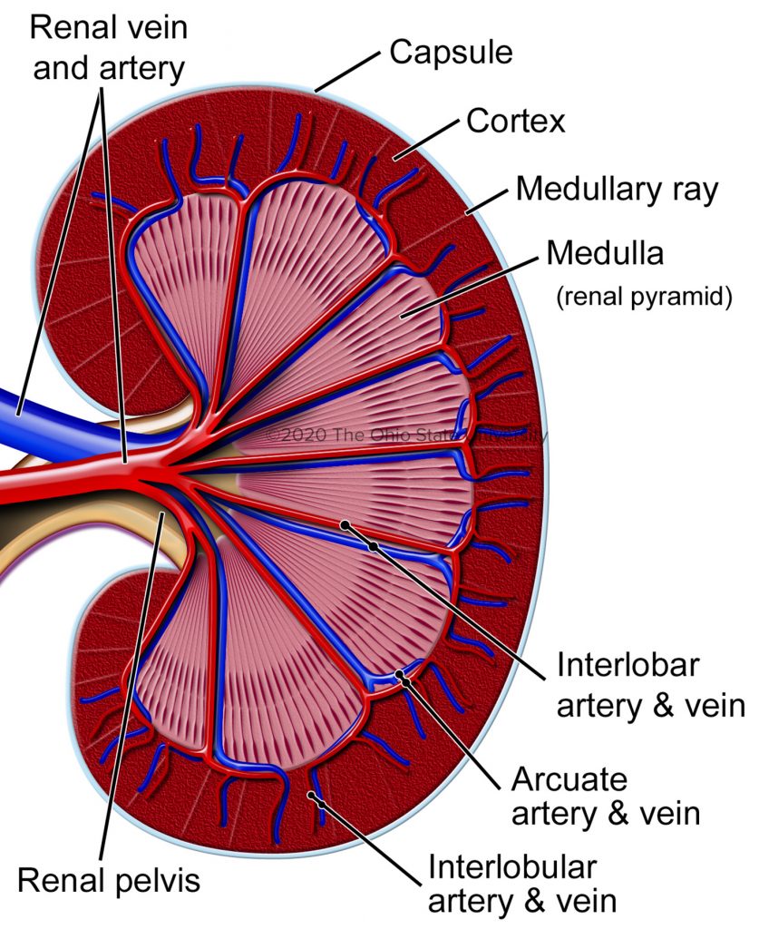
Overview And Anatomy Veterinary Histology from ohiostate.pressbooks.pub These give off a series of branches which enter the cortex as interlobular arterioles. Labeled a in this picture The renal artery enters the kidney via the renal hilum. Fill in the missing blanks for the path of blood flow through the kidney. Label the blood vessels of the kidney in figure 11.5 by filling in the blanks below the figure. The inner portion of each kidney contains a region called the renal medulla. The renal arteries arise directly from the aorta, and the renal veins drain directly into the inferior vena cava. Blood vessel names and roles are explained in this video, beginning with renal artery and ending with the cortical radiate arteries that serve the glomeruli.
Ureters, blood vessels, lymph vessels, and nerves enter and leave at the renal hilum.
There are two blood vessels leading off from the abdominal aorta that go to the kidneys. Physiology and anatomy of blood vessels prepared by dr. Filtered blood leaves the glomerulus via the efferent arteriole, which becomes the interlobular vein. They ultimately end as afferent arterioles, which transport blood into the renal glomerulus for filtration. Blood supply of the kidney: The filtered blood leaves through the renal veins. The interlobar arteries, in turn, branch into arcuate arteries, cortical radiate arteries, and then into. Kidney blood vessels labeled / blood supply to the kidneys anatomy pictures and information. The adrenal (or suprarenal) glands are paired endocrine glands situated over the medial aspect of the upper poles of each kidney. But they are depicted in most pictures and labeled as segmental veins, or not labeled. The renal artery enters through the hilum, which is. From these arterioles branch the afferent arterioles.each afferent arteriole divides into a capillary network. Renalan segmental artery arcuote artery afferent arteriole efferent arteriore figure 11.5:
Label the blood vessels of the kidney in figure 11.5 by filling in the blanks below the figure. Most text books say there are no segmental veins, so they are not in the above blood flow slides. Blood supply of the kidney: Blood supply to the kidney. Structure of blood vessel walls.
Urinary System from medcell.med.yale.edu The renal columns house blood vessels figure 24.3 internal anatomy of the kidney, including the nephron. Identify the anatomical structures of the kidney. Kidney disease, mi, heart failure, stroke and blindness dr. Blood supply to the kidney. Oxygenated blood comes to the kidneys. Filtered blood leaves the glomerulus via the efferent arteriole, which becomes the interlobular vein. Make sure that you understand the functions of these blood vessels (use your textbook as a resource) renal arteries. Moving filtrate from the nephrons to the blood.
Labeled a in this picture
Most text books say there are no segmental veins, so they are not in the above blood flow slides. This will be a smaller amount of blood, since much of the blood would leave the capillaries at the glomerulus and enter the nephron. The interlobar arteries which pass between the renal pyramids, arch around the base of the pyramid as the arcuate. The kidneys are composed of three main sections. The renal artery enters the kidney via the renal hilum. Lymphatic drainage superficial lymphatic vessels form a plexus under the renal capsule (thin layer covering the kidneys) known as the subcapsular lymphatic plexus. Label the blood vessels of the kidney in figure 11.5 by filling in the blanks below the figure. The renal artery enters the hilum of the kidney and divides into a series of smaller vessels. They ultimately end as afferent arterioles, which transport blood into the renal glomerulus for filtration. Identify the anatomical structures of the kidney. Compare the anatomy of the sheep kidney to the human kidney. These are the functional units. Only a light or electron microscope can reveal these structures.
These are the functional units. The renal arteries arise directly from the aorta, and the renal veins drain directly into the inferior vena cava. Identify the anatomical structures of the kidney. Blood supply to the kidney. Ureters, blood vessels, lymph vessels, and nerves enter and leave at the renal hilum.
16 Anatomy And Physiology Models Ideas Anatomy And Physiology Physiology Anatomy from i.pinimg.com Blood circulation into and out of the kidneys is highlighted with colored arrows. Kidney blood vessels labeled / blood supply to the kidneys anatomy pictures and information. These are the functional units. Renal system, in humans, organ system that includes the kidneys, where urine is produced, and the ureters, bladder, and urethra for the passage, storage, and voiding of urine. The renal artery provides the blood flow to the kidney. These give off a series of branches which enter the cortex as interlobular arterioles. Make sure that you understand the functions of these blood vessels (use your textbook as a resource) renal arteries. Kidney function is derived from the actions of about 1.3 million nephrons per kidney;
The other side of the renal sinus, bordering the concave surface of the kidney, opens to the outside through the renal hilus.
Renalan segmental artery arcuote artery afferent arteriole efferent arteriore figure 11.5: Blood supply to the kidney. Kidney blood vessels labeled / blood supply to the kidneys anatomy pictures and information. Ureters, blood vessels, lymph vessels, and nerves enter and leave at the renal hilum. The filtered blood leaves through the renal veins. Fill in the missing blanks for the path of blood flow through the kidney. So the veins highlighted below (here or in other depictions of the kidney. Labeled a in this picture From these arterioles branch the afferent arterioles.each afferent arteriole divides into a capillary network. Capillaries arcuate vein renal vein. They also play a role in regulating important components in the blood. But they are depicted in most pictures and labeled as segmental veins, or not labeled. Label the blood vessels of the kidney in figure 11.5 by filling in the blanks below the figure.
All the blood in the body moves in and out of the kidneys hundreds of times each day—that's about 200 quarts of liquid to be filtered every 24 hours blood vessels labeled. Renal artery afferent arteriole figure 11.5:


0 Komentar